Sunday, June 5, 2016

The Case of Suspected Bilateral Squamous Cell Carcinoma

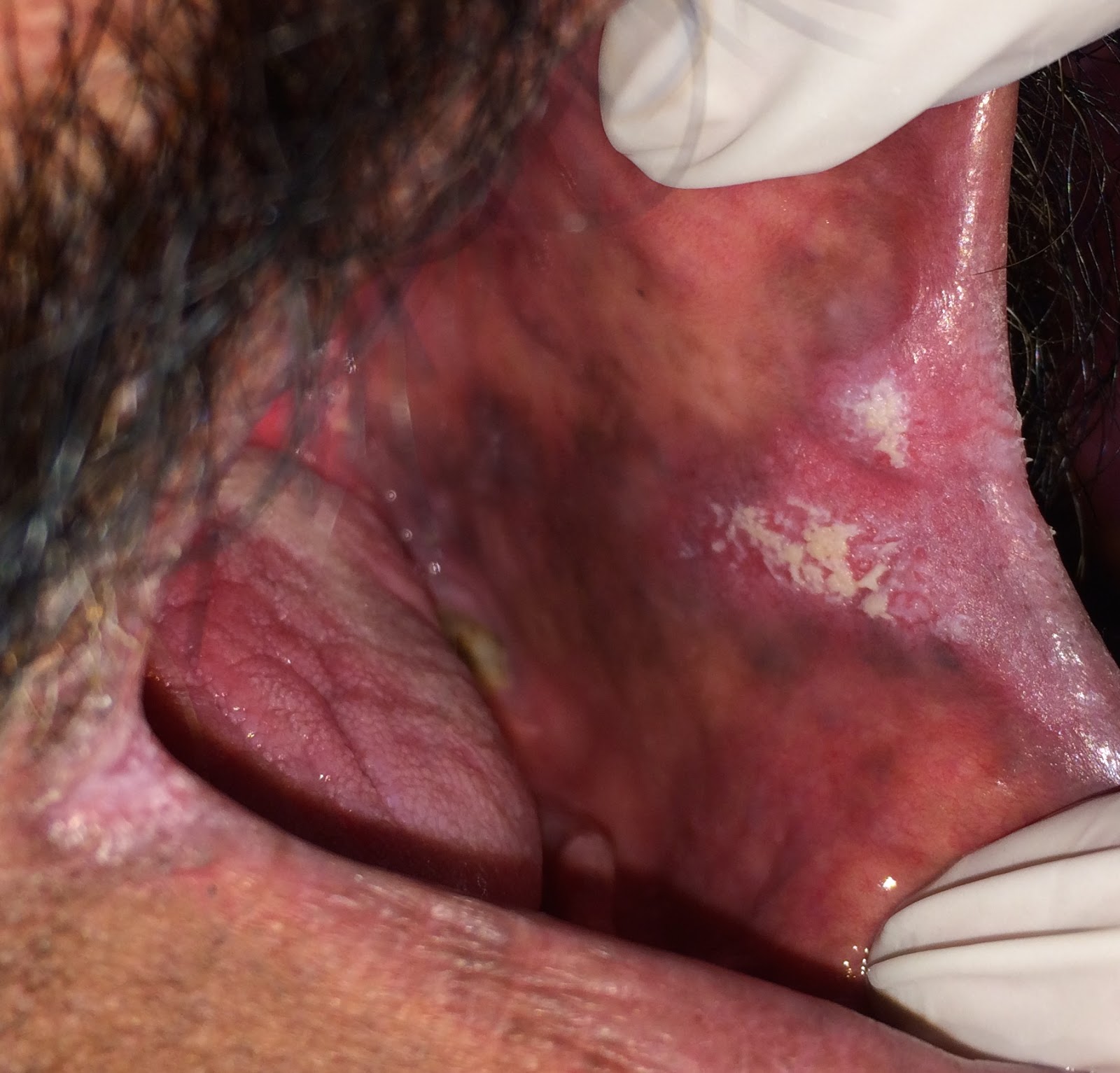
A 48-year-old male reported to the oral surgery department with a complaint of teeth in the lower left side. During the intraoral examination the dental student noted an ulcer in close proximity, the patient told her that the sore had been there for a while but it did not bother him much. Preoperative radiograph of the area showed radiolucency around lower molars. These teeth exhibited grade 3 mobility and were easily extracted. 

Preoperative radiograph of the 48-year-old

The student then went to the clinic supervisor to report that the extractions were complete. During her report she mentioned the ulceration at the site. The clinic supervisor reviewed the patient's history form that was absent of any red flags. She then asked the patient if there was any significant social history. The patient revealed that he had been using smokeless tobacco (niswar in the local language) for several years. Since this side was sore, he was now placing the quid on the opposite side. Intervention at this point was difficult due to the bleeding socket, the patient was instructed to return for a follow up visit in one week's time.

On one week's follow up, the patient presented with a distinct ulcer on the buccal side of the alveolar mucosa.

Ulceration at the site of extraction
A panoramic film was taken. It showed areas of radiolucency bilaterally at the sites of quid placement. The one on the left side was ill-defined and ragged in appearance, highly suggestive of malignancy. The clinic supervisor performed an incisional biopsy at this point. 


Histopathological findings showed a soft tissue specimen consisting of dyspalstic oral epithelium with fibrovascular connective tissue and a malignant neoplasm demonstrating epithelial differentiation. The tumor was invading the underlying connective tissue in nests and cords. The nests exhibited prominent keratin pearl formation in the central areas.

Keratin pearl formation in oral squamous cell carcinoma

Based on the histological findings, a diagnosis of oral squamous cell carcinoma was made.

The patient did not take the news of his cancer well. It took us a while to convince him to let us biopsy the other side. Another incisional biopsy was done in the right mandibular site of placement following extraction of teeth in the region. On histopathology sections, we saw stratified squamous epithelium in association with connective tissue and occasional fragments of bone. The surface oral epithelium showed prominent parakeratinization and what Dr. K calls a "busy" basal layer. The underlying connective tissue showed a chronic inflammatory infiltrate. Dr. K did not like the look of this tissue, she did not think that the cells were maturing appropriately, so she called this tissue atypical and made a comment of close clinical follow up. 

Hyperkeratosis with epithelial atypia, and chronic mucositis
The patient was asked to bring in his imaging studies and was offered partial mandibulectomy with selective neck dissection. He hasn't been back since. If he returns, we might have more images of his resection to share with you. 


Thursday, June 2, 2016

The Case of Aphthous Ulcers

A 21-year-old female dental student reported to the oral surgery department with a complaint of painful ulcers in the mouth. On taking history we discovered that the ulcers appeared 2-3 days ago and she had been in extreme pain since then. The patient told us that the ulcers had appeared once before on the labial mucosa but had healed within a week. Her medical history was clear of any significant medical illness.


21-year-old female with aphthous ulceration on right buccal mucosa

Intraoral examination revealed 2 small ulcers less than 1 cm in size on the buccal mucosa. Both ulcers had a characteristic red halo around it.

Based on the patient's history (spontaneous onset, history of previous ulcers healing in 7 days) and clinical appearance of the lesions (presence on unattached mucosa, distinct red halo around the ulcers), a diagnosis of aphthous ulcers was made. 

Kenalog Orabase gel was prescribed to the patient with directions to apply it 4 to 6 times a day. The patient recovered within a few days.

Notes from Dr. K:
Aphthous ulcers, AKA canker sores, are a common oral pathology with reported incidences of up to 25% in the general population. They fall in the category of autoimmune processes. A variety of etiologies have been proposed as an initiating factor for this process, including allergies, genetics, good disorders, hormonal disorders, nutritional problems, smoking cessation, and following trauma.  Scientific evidence on all these is equivocal. An increased incidence of aphthous ulceration has been reported with systemic diseases, such as nutritional deficiencies, immunodeficiencies, gastrointestinal diseases.

Aphthous ulcers tend to occur in young adults more frequently, especially those who lead high stress lives. The ulcers are classified into three types, minor, major and herpetiform. The case that Dr. M's shared today is an example of the minor type. The ulcers are usually multiple, between 3-10 mm in dimensions, and exclusively involve parakeratinized mucosa (everything except for the alveolar ridge and hard palate). The lesions often appear at one site, heal over a period of 7 days and occur elsewhere. Patients may have one episode in a lifetime, or may not experience a day of relief without the ulcers.

The diagnosis is usually made on clinical grounds, on the basis that Dr. M states with the case above.  Potent topical corticosteroids help in a faster recovery. They also seem to help in decreasing the frequency of ulceration. In patients with continuous episodes, it may be worthwhile to undergo a complete systemic evaluation.

Another case of aphthous ulcers: http://mydayinclinic.blogspot.com/2015/02/the-case-of-two-pathologies-part-1.html


Tuesday, May 31, 2016

The Case of a Blue Lesion on Tongue

A 19-year-old female was seen in the surgery clinics today with a complaint of a blue lesion on her tongue present for as long as she could remember. The lesion was asymptomatic and she did not think that it had increased in size. The patient's medical history was positive for epilepsy. The seizures began a few years ago, but had been decreasing in intensity progressively. She had been taking medication sporadically for her condition. At present she was off all medication on the advice of her doctor. Her concern was whether the blue lesion in her mouth had anything to do with her epileptic seizures.

19-year-old with a hemangioma on dorsal tongue
Intraoral examination was insignificant except for a 12 x 10 mm purplish-blue lesion on anterior dorsal tongue. The lesion was slightly elevated and was relatively firm on palpation.

Based on the long-term history and clinical appearance, a diagnosis of hemangioma was made.

The patient was informed of this diagnosis. She was also told that the hemangioma and her epilepsy were two independent conditions. We offered to remove the hemangioma but the patient decided against it for the moment.

Hemangioma:
Hemangiomas are benign vascular tumors. They are most common in infancy. Females are more commonly affected than males. In infants, hemangiomas rapidly increase in size over a period of time. Following this drastic increase in size, most tumors begin to involute. By age 9, 90% of the lesions have resolved to some degree.

Initial lesions are usually red, older lesions tend to become darker in color. Some hemangiomas are flat and present only as color changes, while others form obvious masses of variable sizes. Bone involvement, although rare, can occur. The classic radiographic appearance of intraosseous hemangiomas is described as a multilocular defect with "honey-comb", or "soap bubble" appearances.

Histologically hemangiomas are classified into various types. Although these histological types usually don't have a significant effect on the prognosis of the lesion, they may effect the treatment plan.

For young children with hemangiomas, periodic observation may the best course of action. If the lesion fails to involute and results in significant esthetic concerns, surgical removal can be attempted. Since this lesion is vascular and there is a risk of significant bleeding, sclerotherapy should be attempted before intervention. 

Tuesday, May 24, 2016

The Case of Multifocal Candidiasis

A 42-year-old male was seen in the Oral Surgery clinics for a tooth extraction. During the oral examination, the student noted red and white changes in the patient's mouth. That's when he decided to call me down for a consult.

The patient was asymptomatic. He was not aware of the presence of lesions inside his mouth. His medical history was insignificant, but he had been a smoker for over 20 years. He did not wear dentures currently.

During the intraoral exam, tiny reddish bumps were noted on the erythematous palate. The tongue exhibited atrophy of the filiform papillae in the central portion and white patches were noted in the anterior buccal mucosa bilaterally. The white patches on the buccal mucosa could not be removed with a piece of dry gauze. 

Inflammatory papillary hyperplasia on the erythematous palate

Central papillary atrophy

Hyperplastic candidiasis in anterior buccal mucosa
Although the clinical features were highly suggestive of candidiasis, a culture was done to establish definitive diagnosis.
Positive candida culture on sabouraud's agar
The patient was prescribed a course of antifungals. Since Clotrimazole troches are not available here in Pakistan, we have to prescribe Nystatin drops. Solutions are a little hard to hold in the mouth for an extended period of time (the solution needs to be held in the mouth for at least 2 minutes for it to be effective). This is a significant factor in noncompliance. 

The patient was instructed to rinse and spit 2 teaspoons of the Nystatin solution five times per day. Unfortunately, the patient never returned for a followup, a dilemma that we commonly encounter with our patients here. It would have been great to include followup pictures. 

Candidiasis:
Candida albicans forms a part of the normal oral flora in approximately 40% of Pakistanis. In situations of immunosuppression, or alteration of the normal floral balance (dentures, antibiotic use, corticosteroid inhaler use etc.), it can cause visible oral changes. 

One of the most common clinical presentation of the infection is the atrophy of filiform papillae in the central portion of the tongue, also known as central papillary atrophy (previously called median rhomboid glossitis). Our patient had a very prominent presentation of this process.

In patients that wear dentures palate involvement is common, especially at the site where the denture comes into contact with the mucosa (denture stomatitis). The redness on the palate may be due to direct inoculation of candida onto the mucosa, or due to the irritation of the palatal mucosa because of the organisms on the denture. The palatal bumps are called inflammatory papillary hyperplasia. These are seen in patients with ill fitting dentures and mouth breathers. They don't necessary have to appear red and inflamed. Superimposition of candida is probably the cause behind the redness. Our patient was a little confused about his denture status but due to the presence of the small red bumps, we suspect that dentures were involved at some point. 

Another cause of palatal involvement is due to transfer of organisms from the tongue to the palate. The infected area becomes erythematous. Such lesions are referred to as kissing lesions. 

White lesions associated with candidiasis commonly rub off when attempts are made to remove them with a piece of dry gauze or a tongue depressor. This form of candidiasis is known as pseudomembranous and was not evident in our patient. White lesions that cannot be removed in a case of candidiasis are called hyperplastic candidiasis or candidal leukoplakia. The most common site of occurrence in anterior buccal mucosa, exactly the site where our patient presented with the lesions. 

The reason as to why this patient developed such a florid presentation of candidiasis remains unknown. It could be due to some medical condition that he's currently unaware of or refuses to inform us of, or it could be simply because of the failure to maintain proper hygiene. 

Saturday, May 21, 2016

The Case of Ameloblastoma

A 58-year-old female patient came to surgery department with a panoramic film that had a well-defined, unicystic radiolucency in the lower right premolar region. The incional biopsy was signed out as ameloblastoma. The surgeons performed an excisional biopsy with extensive curettage and sent the specimen to our service for histopathology. They told us that the clinical presentation was unremarkable.
Radiograph exhibiting a well-defined, unilocular radiolucency in the right premolar region
(Courtesy: Dr. Sidra-tul-Muntaha)
Multiple large nests of odontogenic epithelium with tall, columnar cells at the periphery. Squamous metaplasia can be noted in the central portion of the islands. 
On histopathology, tumor cells appeared to be organized in the form of nests were seen. The cells at the periphery of the nests were tall and columnar and had nuclei exhibiting reverse polarity. Center of the nests were more loosely arranged, resembling stellate reticulum of a developing tooth bud. These features are characteristic of ameloblastoma, more specifically follicular type in this case. Interestingly, a few tumor nests with squamous metaplasia were also noted, a feature that is prevalent in acanthomatous types of ameloblastomas.

High-power view exhibiting reverse polarity in the peripheral tumor cells
 I called this follicular ameloblastoma exhibiting areas of squamous metaplasia.


Dr. K's Comments:
Ameloblastoma:
Ameloblastoma is a benign odontogenic tumor that is seen exclusively in the jaws, because that is the only site where odontogenic epithelium exists. It is seen in individuals over the age of 40 and does not demonstrate any sex predilection. It appears as a painless mass, that can continue to grow to large proportions if not removed timely. Posterior mandibular area is the most common site of involvement.

The radiographic presentation can vary from a well-demarcated, unilocular radiolucency to large multilocular defects. Resorptions of roots in the vicinity of the tumor may also be noted.

Diagnosis can only be confirmed through incisional or excisional biopsy, depending on the size of the lesion. Although the literature describes 6 distinct histological types of ameloblastomas, suggesting that each tumor falls in either one category or the other, there is rarely a tumor that demonstrates less than two histological types. In our case the predominant type was follicular, but we also saw a small area with a plexiform appearance and some tumor islands with acanthomatous change.

The preferred form of management is marginal resection. Simple enucleation followed by curettage can leave some tumor islands behind.


Thursday, May 19, 2016

The Case of Palate Ulcer

A 24-year-old patient came to the department with the complaint of pain in the roof of his mouth. While taking history, I discovered that his pain started a few days after the extraction of the tooth at the site where local anesthesia was administered. Oral examination revealed a well-circumscribed ulcer, about 10 x 5 mm in size, and reddish-yellow in color on the hard palate.


My differential diagnosis included anesthetic necrosis and necrotizing sialometaplasia. The clinical findings, with support from history helped confirm my diagnosis of anesthetic necrosis.                                                                

Notes from Dr. K
Anesthetic Necrosis:
Anesthetic necrosis is usually a result of excess pressure application during local anesthesia administration in attempts to deposit an increased amount of anesthetic solution. Localized ischemia results that presents as a well-circumscribed, deep ulcer. The ulcer heals over a period of time. In persistent lesions a small degree of intervention can encourage healing. As Dr. M says, the history and clinical presentation were highly suggestive of this diagnosis.  

Necrotizing sialometaplasia was easy to rule out in this scenario, the ulcer was on the hard palate (no salivary glands there), there was absence of the classic history of "palate falling off", and the condition usually takes a couple of weeks to evolve from a swelling to a crater (our case was only a few days old judging from the condition of the healing socket). 

Saturday, March 7, 2015

The Case of a Soup Burn

A 27-year-old male was seen in the general practice clinic for a cleaning when the hygienist noticed an area where the superficial surface of the mucosa was peeled off. A blister had appeared at the site following consumption of hot soup a couple of days ago. It had eventually peeled off. The area was only slightly uncomfortable and the patient did not seem particularly worried about it. 

Soup burn in a 27-year-old male
No treatment was recommended on this case.


Thursday, February 26, 2015

The Case of the Yellow Thing in the Eye

A student of mine was curious about the round, yellowish bulge in his patient's eyes.

Pinguela

That small, yellowish bulge on the medial side of the cornea is called a pinguecula. The bulge forms due to the thickening of the conjunctival epithelium. This is a result of constant irritation, usually through excessive exposure to sunlight or dust. The process usually requires no treatment. 

The Case of Leukoplakia

A 39-year-old female was seen in the dental hygiene clinic for a routine cleaning appointment. During the initial exam, the student noticed this white lesion on her posterior buccal gingiva. The patient had first noticed the lesion several months ago. As far as she could tell, there had been no recent changes in size or appearance. 

Intra-oral exam revealed a well-demarcated white lesion on the gingiva below the mandibular canines and premolars. The lesion was approximately 15 x 10 mm in size. The lesion was a little non-homogenized in appearance.

39-year-old female with leukoplakia


Based on the appearance, this lesion was diagnosed as leukoplakia. A biopsy of the lesion was recommended. The periodontist performed an incisional biopsy. The pathologist felt that the tissue appeared pre-neoplastic under the microscope. The case was signed out as mild epithelial dysplasia. The patient was scheduled for regular follow-ups to monitor changes in size, texture and appearance. If any of these was detected, a re-biopsy was strongly recommended.

For a detailed discussion on leukoplakia, please see post, The Case of a White Lesion.

Oral Dysplasia:

Dysplasia is usually a difficult concept to grasp for clinicians. The best way to describe dysplasia is as "pre-cancer". It is a phase that the epithelial cells go through before invading the underlying connective tissue. According the extent of epithelial involvement, dysplasia is classified as mild, moderate and severe.

It is important to keep in mind that the criteria according to which oral dysplasia is classified is very different than the rest of the body. Oral dysplasia tends to be a lot more subtle. General pathologists are more familiar with cervical dysplasia and try to enforce a similar criteria for oral dysplasia, this usually results is misdiagnosis and delayed intervention. 

Thursday, February 19, 2015

The Case of a Foreign Body Tattoo

This 12-year-old boy was undergoing an initial evaluation at the orthodontics department when a grey-colored spot was noted above his left lateral incisor. Since there were no amalgams on his teeth, or had ever been, the orthodontist was curious about what it was.

I asked the mother if the patient had a history of trauma in the region. There had been an a bicycle accident when the boy was 6. He had lost his left central and lateral incisors as the result of it. She remembered that small spot being there for several years but couldn't recall exactly when it had appeared.


12-year-old boy with foreign body tattoo
This was called a foreign body tattoo. The most likely conclusion is that some kind of foreign material got incorporated at the site at the time of the accident. However, a pencil injury (graphite tattoo) or some other incident cannot be completely ruled out. No treatment is usually required for this condition unless the patient has esthetic concerns. In those cases, a simple surgical excision is sufficient.