A 42-year-old male was seen in the Oral Surgery clinics for a tooth extraction. During the oral examination, the student noted red and white changes in the patient's mouth. That's when he decided to call me down for a consult.
The patient was asymptomatic. He was not aware of the presence of lesions inside his mouth. His medical history was insignificant, but he had been a smoker for over 20 years. He did not wear dentures currently.
During the intraoral exam, tiny reddish bumps were noted on the erythematous palate. The tongue exhibited atrophy of the filiform papillae in the central portion and white patches were noted in the anterior buccal mucosa bilaterally. The white patches on the buccal mucosa could not be removed with a piece of dry gauze.
 |
| Inflammatory papillary hyperplasia on the erythematous palate |
 |
| Central papillary atrophy |
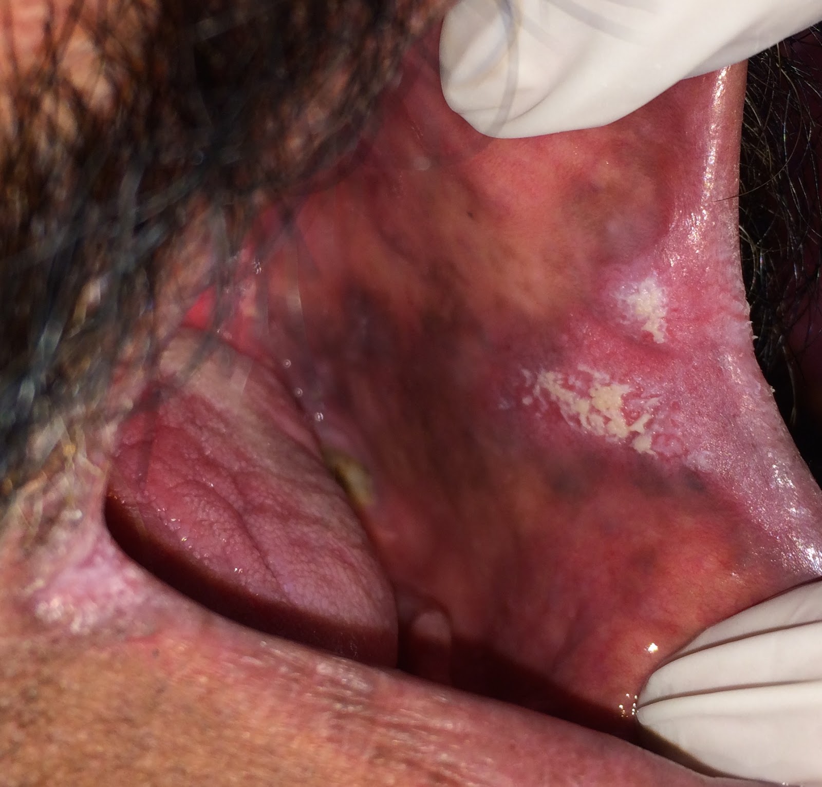 |
| Hyperplastic candidiasis in anterior buccal mucosa |
Although the clinical features were highly suggestive of candidiasis, a culture was done to establish definitive diagnosis.
 |
| Positive candida culture on sabouraud's agar |
The patient was prescribed a course of antifungals. Since Clotrimazole troches are not available here in Pakistan, we have to prescribe Nystatin drops. Solutions are a little hard to hold in the mouth for an extended period of time (the solution needs to be held in the mouth for at least 2 minutes for it to be effective). This is a significant factor in noncompliance.
The patient was instructed to rinse and spit 2 teaspoons of the Nystatin solution five times per day. Unfortunately, the patient never returned for a followup, a dilemma that we commonly encounter with our patients here. It would have been great to include followup pictures.
Candidiasis:
Candida albicans forms a part of the normal oral flora in approximately 40% of Pakistanis. In situations of immunosuppression, or alteration of the normal floral balance (dentures, antibiotic use, corticosteroid inhaler use etc.), it can cause visible oral changes.
One of the most common clinical presentation of the infection is the atrophy of filiform papillae in the central portion of the tongue, also known as central papillary atrophy (previously called median rhomboid glossitis). Our patient had a very prominent presentation of this process.
In patients that wear dentures palate involvement is common, especially at the site where the denture comes into contact with the mucosa (denture stomatitis). The redness on the palate may be due to direct inoculation of candida onto the mucosa, or due to the irritation of the palatal mucosa because of the organisms on the denture. The palatal bumps are called inflammatory papillary hyperplasia. These are seen in patients with ill fitting dentures and mouth breathers. They don't necessary have to appear red and inflamed. Superimposition of candida is probably the cause behind the redness. Our patient was a little confused about his denture status but due to the presence of the small red bumps, we suspect that dentures were involved at some point.
Another cause of palatal involvement is due to transfer of organisms from the tongue to the palate. The infected area becomes erythematous. Such lesions are referred to as kissing lesions.
White lesions associated with candidiasis commonly rub off when attempts are made to remove them with a piece of dry gauze or a tongue depressor. This form of candidiasis is known as pseudomembranous and was not evident in our patient. White lesions that cannot be removed in a case of candidiasis are called hyperplastic candidiasis or candidal leukoplakia. The most common site of occurrence in anterior buccal mucosa, exactly the site where our patient presented with the lesions.
The reason as to why this patient developed such a florid presentation of candidiasis remains unknown. It could be due to some medical condition that he's currently unaware of or refuses to inform us of, or it could be simply because of the failure to maintain proper hygiene.
No comments:
Post a Comment