Tuesday, December 20, 2016

The Case of Herpetic Gingivostomatitis

We were called for a consult at the Orthodontics Clinics for a teenage patient who had developed ulceration on her lips and tongue two days following placement of orthodontic bands. No medications were prescribed to the patient and the patient denied taking any medications during this period. The orthodontist was concerned for erythema multiforme because of the lip involvement. 

We asked the patient a few more questions. The lesions were extremely painful, she hadn't eaten anything for almost 4 days. She had a low grade fever. 

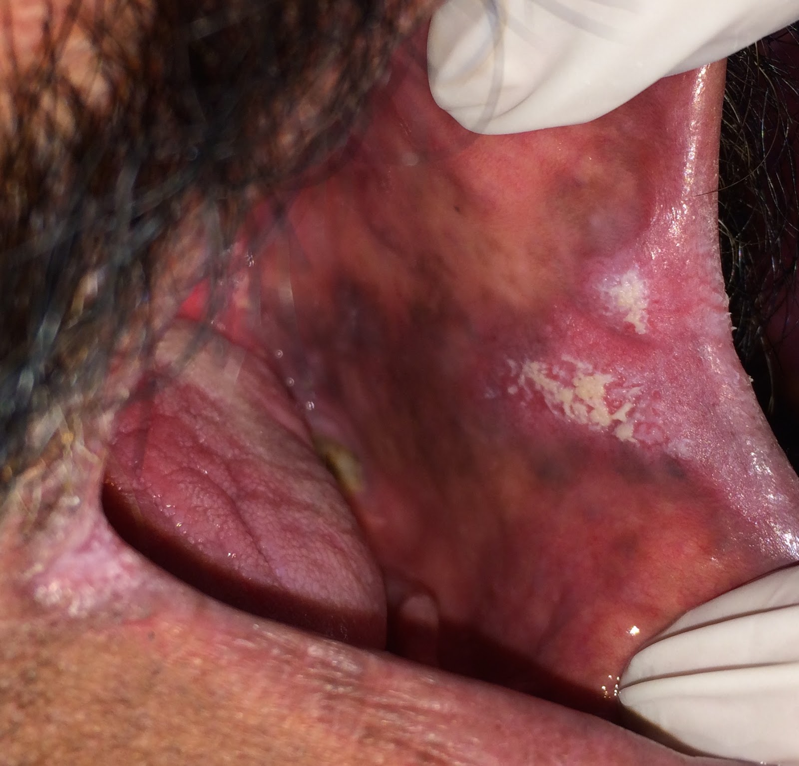
A complete oral examination was difficult because the oral mucosa was extremely tender. We were able to identify a couple of ulcers on the tongue, erythema and swelling on anterior gingiva and a few ruptured blisters on the lips. The lips were slightly swollen. The ulcers were really tiny, about 1-2 mms in size. No other lesions were noted on the body. (Apologies for the bad quality of the pictures, the patient was in a lot of pain and was unable to open her mouth fully for better pictures, she was also reluctant in allowing us to touch her lips, resulting in a very limited view).

A tiny ulcer visible on the tip of the tongue

Ruptured blisters visible on multiple sites on the lips

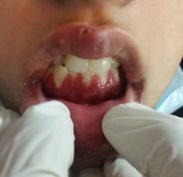
Erythematous, swollen gingiva

Although we could have attempted to perform a cytology on the lip lesions, the patient was already in a lot of pain and not in a mood for letting us intervene in any way. 

We told the patient to get rest, take lots of fluids, consume soft diet and apply topical anesthetic agents for pain relief. It also took us a while to convince her that the infection has nothing to do with her orthodontic treatment. Although she had forced the orthodontist to remove her bands before her consult, she did reluctantly agree to pursue her orthodontic treatment further.

On one week follow-up, the patient's lesions had healed and she was in good health. 

Primary Herpetic Gingivostomatitis:
Herpetic gingivostomatitis is a very common infection caused by the herpes simplex type I virus. The virus spreads through the saliva. Children tend to be more commonly infected because of their habit of putting things in their mouth, somewhere along the way this leads to contact with contaminated saliva and consequently the disease. Unexposed adults can acquire the infection from children. Adult infections tend to be more severe.

The infection is characterized by tiny oral blisters that rupture to form ulcers that are usually 1-2 mms in size. Larger lesions may be encountered but they usually result from multiple small ulcers coalescing. The lesions may involve any oral surface. Involvement of lips is relatively rare, swelling associated with lip involvement is even rarer. 

Once the viral infection has manifested there is very little you can go except for providing supportive care, like we did in this case. If it is diagnosed at an earlier stage, antivirals may be prescribed to limit the course of disease. 

It is important to note that once infected the virus never really leaves your body. It hides away in your nerves and is reactivated in times of stress. Recurrent lesions always appear at the same site, lips being the most common site. Intraoral presentation of recurrent disease is relatively uncommon. The lesions are preceded by a prodrome (itching or tingling) before the appearance of blisters. 


Saturday, December 17, 2016

The Case of the Disappearing Salivary Gland Swelling

A 40-year-old female came to the clinic complaining of a swelling that appeared over her parotid region, was slightly painful but resolved over half an hour. She could speed up the process by applying pressure over the gland. She said that when she applied the pressure, the taste in her mouth became somewhat salty. There were no constitutional symptoms. The patient had seen a couple of dentists before coming to us, she had been advised a series of tests including a panoramic film, ultrasound, complete blood picture and fine needle aspiration cytology. Their listed differential diagnosis was pleomorphic adenoma?

When I examined the patient there was no identifiable swelling, both extra and intra-orally. Thick, mucinous saliva was expelled from the salivary gland on initial milking. The saliva on persistent milking was watery. The panoramic film was clear of any obvious pathology, the ultrasound report was suggestive of inflammation in the salivary gland, and the blood work was normal. Since there was no identifiable pathology, the pathologist had refused to perform an FNAC. 

So we concluded that there was some blockage in the salivary gland duct that was preventing the salivary gland duct to drain completely resulting in the swelling. Once the duct was able to drain the saliva, the swelling would disappear. The blockage in the duct was obviously not because of a lith, we would have been able to see that on radiograph. The cause was most likely a mucus plug. 

Conservative management required us to push the mucus plug out by increasing the salivary flow. We recommended compression of the gland at least twice daily and use of sugar-free lemon lozenges multiple times a day. The patient called after 4 days saying that she was symptom free, and wanted to discontinue lemon lozenges because she didn't care much for the taste. We asked her to complete 7 days and then let us know how she's feeling. 

She continues to be symptom-free for about 2 weeks now. Whatever was obstructing the duct was expelled out by the salivary gland stimulation. 

Comments:
Swellings that grow and regress in size over a period of time are usually inflammatory in nature. Pleomorphic adenoma is a benign salivary gland tumor and was not reasonable differential here. Neoplastic growths don't regress, instead they continue to increase in size. 

Also pleomorphic adenoma would be palpable. No palpable growth was identified during the oral examination. 


Monday, November 28, 2016

The Case of Weird-Looking Tongue

A 30-year-old female patient came to the surgery department for extraction of her tooth. The student dealing with the patient noticed this "weird looking pattern" on the tongue. She called us for a consult. The patient was not aware of her condition, had never experienced any symptoms associated with this. Intraoral examination revealed red patches surrounded by a distinct white outline. The central redder areas appeared to be depapillated. 

Geographic tongue in a 30-year-old female
The condition was diagnosed as geographic tongue, based on the characteristic clinical appearance. 
More details about geographic tongue can be found at  the following link: http://mydayinclinic.blogspot.com/2015/02/the-case-of-two-pathologies-part-2.html

Saturday, November 26, 2016

The Case of Trigeminal Neuralgia

An older female patient reported to the Oral Surgery clinics with a complaint of electric-shock like pain on the facial region. She pointed to a region on the face where any contact would elicit the pain. It was on right cheek. The pain lasted for a few seconds. The patient had extracted the teeth in that site over a period of time. She had a stroke a couple of years ago and had paralysis on the left side of the face and weakness in the upper limb. She was using medication for hypertension and was using anticoagulants to prevent future strokes. She was severely disturbed by her symptoms.

The condition was rightly diagnosed as trigeminal neuralgia by her physician in a different city who gave her 400 mg of Carbamezapine (200 in the morning and 200 before bedtime). Although the medication helped relieve her symptoms, the side effect of dizziness became intolerable, so she had to stop using the medication. 

When she came to me the surgeon was already contemplating temporarily anesthetizing the infraorbital nerve through a glycerol injection. Another doctor had suggested using 75 mg of Lyrica (Pregabalin) twice a day. She tolerated the medication well but called me two days later saying that the pain was still there. Another 75 mgs was added in the afternoon and the patient was asked to give the medication at least a week to start acting. She called the next day saying that her symptoms were under control. The patient is continuing this regimen. She follows up 2-monthly by a personal visit or through phone call. 

Trigeminal Neuralgia:
Trigeminal neuralgia is a type of neuropathic pain, which means that the nerve is messed up somewhere along its path resulting in the pain sensation. The cause of the damage to the nerve is usually unidentifiable. The criteria for diagnosis is fairly simple, electric-shock like pain, along the course of the trigeminal nerve, elicited on contact with a "trigger zone". All three of these features were reported in the case we discuss above. 

Although any three of the trigeminal branches can be affected, the maxillary branch is most commonly involved, like in our case. The affected population is usually comprised of individuals over the age of 50. 

Because the initial symptoms may be more sporadic, the condition is usually misdiagnosed as an odontogenic problem. This results in most patients going through repeated endodontic therapy and extractions at the site of pain. Once the classic symptoms set in and the patient is seen by an oral pathologist, oral medicine expert or a neurologist, the teeth in the vicinity of the pain are all missing. We witnessed this in our patient. 

Management of the disease can be complex. It is important to inform the patient that the drugs for this condition work differently than other analgesics. The pain will not stop in a few hours, it may take up to a week for the medication to begin working. It is also important to start from the minimal dose up, for Carbamezapine, the most popular drug used for management of this process, it is 200 mg (100 in the morning and 100 before bedtime). Other medications that can be used are Pregabalin, Gabapentin and Phenytoin. When the medications fail to work, we have to rely on surgical options. 

Glycerol injections are sometimes used to temporarily anesthetize the nerve at the site where it exits the skull. It is a risky procedure because it can result in uncontrolled damage in the area. It may also cause only partial damage to the nerve causing dysaesthesia. The effects of the injection last about 3-6 months. So this is not something to favor over medical therapy. 

Other surgical procedures include microvascular decompression of vessels impinging the nerve in the skull, application of heat to sever the nerve through gamma knife procedure and neurectomy. The success of these procedures varies between 70-80%. 


Monday, September 5, 2016

The Case of Peripheral Ossifying Fibroma

An 18-year-old female patients came to the OMFS department with complaint of discomfort on chewing. Oral examination revealed a pinkish-red colored lesion on buccal and lingual gingiva in the interdental area of mandibular central incisors. Lesion was soft in consistency and measured approximately 2 cm in diameter. Patient said that it's been there for two months.

Excisional biopsy was performed by the oral surgeon. 

Considering the gingival location, the top entities in our differential diagnosis were pyogenic granuloma, peripheral ossifying fibroma and peripheral giant cell granuloma.


Microscopic examination showed a nodule of mucosa surfaced by stratified squamous epithelium. The body of nodule was composed of benign proliferation of plump fibroblasts. Bone formation is identified in some portions. Based on this histopathological presentation, a diagnosis of peripheral ossifying fibroma was made.

Histological slide showing plump fibroblasts and calcifications


Peripheral Ossifying Fibroma:
The peripheral ossifying fibroma is a benign proliferation that occurs exclusively on the gingiva. It is seen more commonly in teenagers and young adults. It usually presents as a pedunculate or sessile nodular mass in the interdental region clinically. The color of the nodule ranges from red to pink depending on the degree of irritation.
Conservative surgical excision is the preferred form of management for this process. Despite their benign nature, these lesions have the ability to become pretty large. 

Sunday, September 4, 2016

The Case of a Huge Mandibular Radiolucency

A 35-year-old patient came to the department with the complaint of pain and swelling in lower left region for the past one year. The swelling was smaller initially but had been gradually increasing in size. The rate of growth had accelerated in the past couple of months. The complaint of pain associated with this swelling was relatively recent. 

On oral examination an ulcerated lesion was noted on the lower left alveolar ridge  The swelling was tender on touching and firm on palpation. No palpable lymph nodes were identified during the extra oral examination. The patient's medical history was significant for a "back" problem. She was wheel chair bound for that purpose.

Clinical picture showing the ulcerated swelling on the left alveolar ridge extending to the buccal vestibule
A multilocular radiolucency was identified in the radiograph pushing the impacted third molar against the border of mandible. In some places a sclerotic margin could be identified at the margins of the radiolucency.

OPG showing the multilocular radiolucency in association with an impacted third molar
The surgeons performed an intraoral biopsy of the lesion from the alveolar ridge. The tissue was somewhat jelly-like in texture.
Biopsy specimen of the patient


The pathologist signed out the case as an odontogenic myxoma. Her report indicated that the sections showed a benign tumor with loose, myxomatous appearance. The cells were spindle shaped and fine collagen fibrils could be appreciated. 

The patient was scheduled for surgery but during her medical evaluation a murmur was identified and the anesthesiologist refused to put her under. We hope that that her medical issue is resolved so she can be treated for her disease.

Dr. K's Comments:
Odontogenic myxomas are rare, benign odontogenic tumors. They tend to favor the posterior mandibular area of young people, the average age is between 25 and 30. Smaller lesions may present as unilocular radiolucencies, while larger lesions, like ours are predominantly multilocular.

Some oral pathologists believe that odontogenic myxomas, odontogenic fibromyxomas and odontogenic fibromas are a spectrum of the same process. The classification varies depending on the amount of identifiable collagen. Since this process is exclusively found in the jaws, general pathologists usually end up lumping it in the fibromatosis category. Since both processes are benign and aggressive, I suppose even the incorrect diagnosis doesn't affect the prognosis. 

This process is managed through surgery. Since the lesion is not encapsulated, its cells infiltrate into the surrounding bone making definition of margins really difficult. Aggressive curettage following removal of the tumor is usually done to ensure complete removal. Periodic re-evaluation is also necessary to monitor for recurrences.

Saturday, August 27, 2016

The Case of a Large Cystic Cavity in the Mandible

A 17-year-old patient came to the oral surgery department with a swelling on lower left side of mandible. The swelling was first noticed one year back, it has slowly progressed in size since then. On clinical examination, a bony hard swelling was palpable in the lower left buccal vestibule intraorally and anterior to the angle of mandible extraorally. The lymph nodes were not palpable.

Radiographic examination showed a well-circumscribed radiolucency in the body of mandible,  involving the roots of left mandibular molars and premolars. A large carious defect was visible in the mandibular first molar, distal root of involved second molar showed a some degree of resorption.

Radiograph shows well demarcated radiolucent area involving roots of mandibular molars and premolars
The radiographic differential was extensive including odontogenic cysts, tumors, and other benign entities. 

When the surgeon aspirated the cavity, a bloody aspirate was noted. She still decided to go in for an incisional biopsy. During the procedure she aspirated more than 15 ml of bloody fluid. She was able to scrape off some tissue from the bone margins. From her description of an empty cavity with almost no tissue and her having to scrape off tissue from the bone margins, we were favoring a diagnosis of traumatic bone cyst. However, the resorption of teeth didn't fit.

The histopathology results were quite surprising.

Histopathological slide showing epithelial lining, granulation tissue and chronic inflammatory cells

Histopathological examination showed granulation tissue partially lined by non-keratinized stratified squamous epithelium. The granulation tissue supported a moderate infiltrate of predominantly chronic inflammatory cells. Based on these results, a definitive diagnosis of radicular cyst (periodical cyst) was made.

Dr. K's Comments:
Radicular Cysts:
The surgeons were as surprised by this presentation as we were. They extracted the tooth, inserted asdrain and asked the patient to return for followup in two weeks. The patient's father insisted that another biopsy be performed at that time, and this time he wanted his pathologist to look at it. His pathologist did a lot of unrequired immunohistochemistry to determine that we were right the first time. His bone is growing back and once the cavity is sufficiently marsupialized, they will excise it.

Although radicular cysts are very common in the jaws, a presentation like this is odd. They usually don't attain such large sizes, or contain so much bloody fluid. Resorption of tooth roots has been reported but it is rare.


Monday, July 11, 2016

The Cases of Bitter Taste, Painful Mouth, Rough Gingiva and Spiked Tongue

A 48-year-old male patient reported to the department of oral medicine for a complaint of bitter taste in his mouth for over two years. He said that his mouth tasted bitter all the time, constantly making him feel nauseated. He was unable to appreciate the difference between sweet, salty or spicy anymore. There were also minor complaints of his gingiva feeling rough and slight burning sensation on the labial mucosa. The patient had seen several doctors with this problem, had all sorts of investigations done but nothing appeared to be wrong. He was currently taking antidepressants. The intraoral examination was unremarkable. 

We also had a 54-year-old female come in the same week with a complaint of inability to tolerate chillies in food. She said that even black pepper made her buccal mucosa and tongue hurt. These symptoms were not constant, she would be able to enjoy a good meal occasionally. She also said that eating fruits (like mangoes and watermelon) make her tongue feel like it had spikes. She was diagnosed with erosive lichen planus by a general dentist and given a course of topical corticosteroids but her symptoms had not improved. However, it is important to note that her oral examination was unremarkable as well.

We diagnosed both these cases as Burning Mouth Syndrome. 

Patient no. 1 was a little reluctant in accepting our diagnosis. He kept returning for follow ups asking us to give him something that will make his symptoms go away. We gave him 0.25 mg of Clonazepam to take before bedtime on his second visit because there is some evidence in the literature that favors this practice. Upped the dose to 0.5 mg on his third visit, but unfortunately it did not improve his symptoms. The patient was lost to follow up after that. 

Patient no. 2 took her diagnosis relatively well. Once she understood what she had, she refused treatment. She said that now that she knows that it is not something significant, it will not affect her as much. We asked her to return in case she wanted us to try Clonazepam for her symptoms, we have not seen her back since. 

Dr. K's Notes:
Burning Mouth Syndrome:
Burning mouth syndrome has diverse presentations as evident from our cases above. Patients always present with elaborate symptoms that are significantly affecting their lives, but nothing is ever identified clinically. Since dentists and other physicians are usually not aware of this conditions, these patients can go undiagnosed for several years. This just adds to the patient's frustrations. 

In simple words burning mouth syndrome can be explained as nerves relaying the wrong information to your brain. This disturbance can manifest in three ways; pain, altered sensation and modified taste. For example, your nerves are telling your brain that your tongue is sore, the food is bitter and your mouth is feeling rough. At any time, one, two or all three disturbances may be present. 

Unfortunately this is not communicated by the name given to condition. The misnomer gives dentists and other physicians a false impression that "burning" of the tongue is a mandatory symptom. As you can see from our examples, it is not always the case.

The problem arises when we are asked to treat this condition. We can't see nerves, there is no technology that enables us to do this yet. So if we can't see where the damage is, we can't fix it. Most of the treatment modalities tried and tested in literature have failed. A double-blind placebo-controlled study showed promising results with Clonazepam, but only in patients that had the pain component. It is did not treat their pain but did put their symptoms in the background. So in patient no. 1 this was not a practical choice, but we tried it anyways. Patient no. 2 might have been a right candidate for this but she was just ecstatic about the fact that she did not have cancer.

Wednesday, June 15, 2016

The Case of Herpes Simplex Virus

A 22-year-old female presented to the department with a complaint of small, painful ulcers on her gums and the inside of her lips. The ulcers had appeared as small bumps that had ruptured to form ulcers. Constitutional signs were absent. As far as she could remember, this was her first episode of anything like this. 

During her intraoral examination, we noted small ulcers on the gums and labial mucosa.They were small in size approximately 1 to 2mm with reddish appearance. A few of them appeared to have coalesced together and seemed bigger than the rest.
Herpetic ulcers on the gingiva and labial mucosa
Photo credit: Dr Amber Kiyani

Multiple tiny small ulcers, a history of "tiny bumps" rupturing into ulcers supported a diagnosis of herpetic stomatitis. To help confirm this diagnosis, a cytology was done. The results were consistent with a herpetic infection. 


Cytology fo herpes simplex virus
Photo credit: Dr Amber Kiyani

Since the infection had already manifested, we did not prescribe antiviral therapy, we just recommended topical anesthetic agents to help with the pain. The patient was seen on followup 7 days later. The lesions had cleared up until then. 


Saturday, June 11, 2016

The Case of Fibrous Dysplasia Encroaching the Orbital Floor

A 47-year-old female was seen in the oral surgery clinics with a complaint of vision and hearing disturbances that had developed over the past few months. The patient had a diagnosis of fibrous dysplasia from several years ago. She was offered surgical trimming of the lesion at the time of diagnosis, which she refused. 

Slight asymmetry in a 47-year-old patient with fibrous dysplasia
Only slight facial asymmetry was noted during the extra-oral exam. A bony hard bulge was palpable in the left buccal vestibule. It was not prominent enough to be photographed. The patient was requested to return with imaging studies. 

CT scan of a patient with fibrous dysplasia
(Courtesy Dr. Asma Pervez)
On her CT scan, a radio-opaque mass was noted to obliterate the maxillary sinus and encroach the nasal wall and the orbital floor. 

The oral surgeons in the clinic offered the patient hemimaxillectomy. They believed that since the disease process was active, and continued to involve more tissue, it was best to remove it completely. The defect would be filled with a specially designed obturator. This would obviously result in suboptimal esthetics, much worse than what the patient was presenting with. However, the patient's sight would be preserved, and future complications, such as involvement of the base of skull, would be prevented. 

As extreme as this sounds, this was the best approach in my opinion. However, I did speak to the surgeons about the possibility of a relatively conservative plan, possibly removing the orbital plate and contouring the lesion involving the maxillary sinus. They said leaving any part of this lesion would be asking for recurrence.

The patient was not happy with this treatment plan. She hasn't returned to schedule surgery.

Fibrous Dysplasia:
Fibrous dysplasia is a developmental condition that results in replacement of normal bone with fibro-osseous tissue. It is linked to a mutation in GNAS 1 (guanine nucleotide-binding protein), which plays an important role in development of bone, endocrine system and skin pigmentation. The severity of bone involvement depends on the time when the mutation takes place, mutations during early embryonic development can cause multiple bone involvement. 

Fibrous dysplasia is broadly classified into polyostotic (multiple bone involvement) and monostotic (one bone involvement) types. 

Monostotic type is more common. The lesions present as a slow-growing, painless swellings. Patients are usually in their teens at the time of diagnosis. Jaws are a frequent site of involvement. The maxilla is more commonly affected than the mandible. For fibrous dysplasia of the maxilla, the use of the term craniofacial fibrous dysplasia is preferred. It is not uncommon for bones in the vicinity to get involved. 

Radiographically, the lesions of fibrous dysplasia are described as having a "ground glass" appearance.   The margins of the lesion are ill-defined and merge with the adjacent normal bone. Diagnosis is confirmed by biopsy. 

In most cases, fibrous dysplasia stabilizes once the skeletal growth ceases. Our case is an obvious exception. Surgical contouring is done to correct minor asymmetries. Rate of recurrence after the procedure vary between 25-50%. Larger lesions may require surgical resection. Bisphosphonate therapy helps to control polyostotic disease. 

Sunday, June 5, 2016

The Case of Suspected Bilateral Squamous Cell Carcinoma

A 48-year-old male reported to the oral surgery department with a complaint of teeth in the lower left side. During the intraoral examination the dental student noted an ulcer in close proximity, the patient told her that the sore had been there for a while but it did not bother him much. Preoperative radiograph of the area showed radiolucency around lower molars. These teeth exhibited grade 3 mobility and were easily extracted. 

Preoperative radiograph of the 48-year-old

The student then went to the clinic supervisor to report that the extractions were complete. During her report she mentioned the ulceration at the site. The clinic supervisor reviewed the patient's history form that was absent of any red flags. She then asked the patient if there was any significant social history. The patient revealed that he had been using smokeless tobacco (niswar in the local language) for several years. Since this side was sore, he was now placing the quid on the opposite side. Intervention at this point was difficult due to the bleeding socket, the patient was instructed to return for a follow up visit in one week's time.

On one week's follow up, the patient presented with a distinct ulcer on the buccal side of the alveolar mucosa.

Ulceration at the site of extraction
A panoramic film was taken. It showed areas of radiolucency bilaterally at the sites of quid placement. The one on the left side was ill-defined and ragged in appearance, highly suggestive of malignancy. The clinic supervisor performed an incisional biopsy at this point. 


Histopathological findings showed a soft tissue specimen consisting of dyspalstic oral epithelium with fibrovascular connective tissue and a malignant neoplasm demonstrating epithelial differentiation. The tumor was invading the underlying connective tissue in nests and cords. The nests exhibited prominent keratin pearl formation in the central areas.

Keratin pearl formation in oral squamous cell carcinoma

Based on the histological findings, a diagnosis of oral squamous cell carcinoma was made.

The patient did not take the news of his cancer well. It took us a while to convince him to let us biopsy the other side. Another incisional biopsy was done in the right mandibular site of placement following extraction of teeth in the region. On histopathology sections, we saw stratified squamous epithelium in association with connective tissue and occasional fragments of bone. The surface oral epithelium showed prominent parakeratinization and what Dr. K calls a "busy" basal layer. The underlying connective tissue showed a chronic inflammatory infiltrate. Dr. K did not like the look of this tissue, she did not think that the cells were maturing appropriately, so she called this tissue atypical and made a comment of close clinical follow up. 

Hyperkeratosis with epithelial atypia, and chronic mucositis
The patient was asked to bring in his imaging studies and was offered partial mandibulectomy with selective neck dissection. He hasn't been back since. If he returns, we might have more images of his resection to share with you. 


Thursday, June 2, 2016

The Case of Aphthous Ulcers

A 21-year-old female dental student reported to the oral surgery department with a complaint of painful ulcers in the mouth. On taking history we discovered that the ulcers appeared 2-3 days ago and she had been in extreme pain since then. The patient told us that the ulcers had appeared once before on the labial mucosa but had healed within a week. Her medical history was clear of any significant medical illness.


21-year-old female with aphthous ulceration on right buccal mucosa

Intraoral examination revealed 2 small ulcers less than 1 cm in size on the buccal mucosa. Both ulcers had a characteristic red halo around it.

Based on the patient's history (spontaneous onset, history of previous ulcers healing in 7 days) and clinical appearance of the lesions (presence on unattached mucosa, distinct red halo around the ulcers), a diagnosis of aphthous ulcers was made. 

Kenalog Orabase gel was prescribed to the patient with directions to apply it 4 to 6 times a day. The patient recovered within a few days.

Notes from Dr. K:
Aphthous ulcers, AKA canker sores, are a common oral pathology with reported incidences of up to 25% in the general population. They fall in the category of autoimmune processes. A variety of etiologies have been proposed as an initiating factor for this process, including allergies, genetics, good disorders, hormonal disorders, nutritional problems, smoking cessation, and following trauma.  Scientific evidence on all these is equivocal. An increased incidence of aphthous ulceration has been reported with systemic diseases, such as nutritional deficiencies, immunodeficiencies, gastrointestinal diseases.

Aphthous ulcers tend to occur in young adults more frequently, especially those who lead high stress lives. The ulcers are classified into three types, minor, major and herpetiform. The case that Dr. M's shared today is an example of the minor type. The ulcers are usually multiple, between 3-10 mm in dimensions, and exclusively involve parakeratinized mucosa (everything except for the alveolar ridge and hard palate). The lesions often appear at one site, heal over a period of 7 days and occur elsewhere. Patients may have one episode in a lifetime, or may not experience a day of relief without the ulcers.

The diagnosis is usually made on clinical grounds, on the basis that Dr. M states with the case above.  Potent topical corticosteroids help in a faster recovery. They also seem to help in decreasing the frequency of ulceration. In patients with continuous episodes, it may be worthwhile to undergo a complete systemic evaluation.

Another case of aphthous ulcers: http://mydayinclinic.blogspot.com/2015/02/the-case-of-two-pathologies-part-1.html


Tuesday, May 31, 2016

The Case of a Blue Lesion on Tongue

A 19-year-old female was seen in the surgery clinics today with a complaint of a blue lesion on her tongue present for as long as she could remember. The lesion was asymptomatic and she did not think that it had increased in size. The patient's medical history was positive for epilepsy. The seizures began a few years ago, but had been decreasing in intensity progressively. She had been taking medication sporadically for her condition. At present she was off all medication on the advice of her doctor. Her concern was whether the blue lesion in her mouth had anything to do with her epileptic seizures.

19-year-old with a hemangioma on dorsal tongue
Intraoral examination was insignificant except for a 12 x 10 mm purplish-blue lesion on anterior dorsal tongue. The lesion was slightly elevated and was relatively firm on palpation.

Based on the long-term history and clinical appearance, a diagnosis of hemangioma was made.

The patient was informed of this diagnosis. She was also told that the hemangioma and her epilepsy were two independent conditions. We offered to remove the hemangioma but the patient decided against it for the moment.

Hemangioma:
Hemangiomas are benign vascular tumors. They are most common in infancy. Females are more commonly affected than males. In infants, hemangiomas rapidly increase in size over a period of time. Following this drastic increase in size, most tumors begin to involute. By age 9, 90% of the lesions have resolved to some degree.

Initial lesions are usually red, older lesions tend to become darker in color. Some hemangiomas are flat and present only as color changes, while others form obvious masses of variable sizes. Bone involvement, although rare, can occur. The classic radiographic appearance of intraosseous hemangiomas is described as a multilocular defect with "honey-comb", or "soap bubble" appearances.

Histologically hemangiomas are classified into various types. Although these histological types usually don't have a significant effect on the prognosis of the lesion, they may effect the treatment plan.

For young children with hemangiomas, periodic observation may the best course of action. If the lesion fails to involute and results in significant esthetic concerns, surgical removal can be attempted. Since this lesion is vascular and there is a risk of significant bleeding, sclerotherapy should be attempted before intervention. 

Tuesday, May 24, 2016

The Case of Multifocal Candidiasis

A 42-year-old male was seen in the Oral Surgery clinics for a tooth extraction. During the oral examination, the student noted red and white changes in the patient's mouth. That's when he decided to call me down for a consult.

The patient was asymptomatic. He was not aware of the presence of lesions inside his mouth. His medical history was insignificant, but he had been a smoker for over 20 years. He did not wear dentures currently.

During the intraoral exam, tiny reddish bumps were noted on the erythematous palate. The tongue exhibited atrophy of the filiform papillae in the central portion and white patches were noted in the anterior buccal mucosa bilaterally. The white patches on the buccal mucosa could not be removed with a piece of dry gauze. 

Inflammatory papillary hyperplasia on the erythematous palate

Central papillary atrophy

Hyperplastic candidiasis in anterior buccal mucosa
Although the clinical features were highly suggestive of candidiasis, a culture was done to establish definitive diagnosis.
Positive candida culture on sabouraud's agar
The patient was prescribed a course of antifungals. Since Clotrimazole troches are not available here in Pakistan, we have to prescribe Nystatin drops. Solutions are a little hard to hold in the mouth for an extended period of time (the solution needs to be held in the mouth for at least 2 minutes for it to be effective). This is a significant factor in noncompliance. 

The patient was instructed to rinse and spit 2 teaspoons of the Nystatin solution five times per day. Unfortunately, the patient never returned for a followup, a dilemma that we commonly encounter with our patients here. It would have been great to include followup pictures. 

Candidiasis:
Candida albicans forms a part of the normal oral flora in approximately 40% of Pakistanis. In situations of immunosuppression, or alteration of the normal floral balance (dentures, antibiotic use, corticosteroid inhaler use etc.), it can cause visible oral changes. 

One of the most common clinical presentation of the infection is the atrophy of filiform papillae in the central portion of the tongue, also known as central papillary atrophy (previously called median rhomboid glossitis). Our patient had a very prominent presentation of this process.

In patients that wear dentures palate involvement is common, especially at the site where the denture comes into contact with the mucosa (denture stomatitis). The redness on the palate may be due to direct inoculation of candida onto the mucosa, or due to the irritation of the palatal mucosa because of the organisms on the denture. The palatal bumps are called inflammatory papillary hyperplasia. These are seen in patients with ill fitting dentures and mouth breathers. They don't necessary have to appear red and inflamed. Superimposition of candida is probably the cause behind the redness. Our patient was a little confused about his denture status but due to the presence of the small red bumps, we suspect that dentures were involved at some point. 

Another cause of palatal involvement is due to transfer of organisms from the tongue to the palate. The infected area becomes erythematous. Such lesions are referred to as kissing lesions. 

White lesions associated with candidiasis commonly rub off when attempts are made to remove them with a piece of dry gauze or a tongue depressor. This form of candidiasis is known as pseudomembranous and was not evident in our patient. White lesions that cannot be removed in a case of candidiasis are called hyperplastic candidiasis or candidal leukoplakia. The most common site of occurrence in anterior buccal mucosa, exactly the site where our patient presented with the lesions. 

The reason as to why this patient developed such a florid presentation of candidiasis remains unknown. It could be due to some medical condition that he's currently unaware of or refuses to inform us of, or it could be simply because of the failure to maintain proper hygiene. 

Saturday, May 21, 2016

The Case of Ameloblastoma

A 58-year-old female patient came to surgery department with a panoramic film that had a well-defined, unicystic radiolucency in the lower right premolar region. The incional biopsy was signed out as ameloblastoma. The surgeons performed an excisional biopsy with extensive curettage and sent the specimen to our service for histopathology. They told us that the clinical presentation was unremarkable.
Radiograph exhibiting a well-defined, unilocular radiolucency in the right premolar region
(Courtesy: Dr. Sidra-tul-Muntaha)
Multiple large nests of odontogenic epithelium with tall, columnar cells at the periphery. Squamous metaplasia can be noted in the central portion of the islands. 
On histopathology, tumor cells appeared to be organized in the form of nests were seen. The cells at the periphery of the nests were tall and columnar and had nuclei exhibiting reverse polarity. Center of the nests were more loosely arranged, resembling stellate reticulum of a developing tooth bud. These features are characteristic of ameloblastoma, more specifically follicular type in this case. Interestingly, a few tumor nests with squamous metaplasia were also noted, a feature that is prevalent in acanthomatous types of ameloblastomas.

High-power view exhibiting reverse polarity in the peripheral tumor cells
 I called this follicular ameloblastoma exhibiting areas of squamous metaplasia.


Dr. K's Comments:
Ameloblastoma:
Ameloblastoma is a benign odontogenic tumor that is seen exclusively in the jaws, because that is the only site where odontogenic epithelium exists. It is seen in individuals over the age of 40 and does not demonstrate any sex predilection. It appears as a painless mass, that can continue to grow to large proportions if not removed timely. Posterior mandibular area is the most common site of involvement.

The radiographic presentation can vary from a well-demarcated, unilocular radiolucency to large multilocular defects. Resorptions of roots in the vicinity of the tumor may also be noted.

Diagnosis can only be confirmed through incisional or excisional biopsy, depending on the size of the lesion. Although the literature describes 6 distinct histological types of ameloblastomas, suggesting that each tumor falls in either one category or the other, there is rarely a tumor that demonstrates less than two histological types. In our case the predominant type was follicular, but we also saw a small area with a plexiform appearance and some tumor islands with acanthomatous change.

The preferred form of management is marginal resection. Simple enucleation followed by curettage can leave some tumor islands behind.


Thursday, May 19, 2016

The Case of Palate Ulcer

A 24-year-old patient came to the department with the complaint of pain in the roof of his mouth. While taking history, I discovered that his pain started a few days after the extraction of the tooth at the site where local anesthesia was administered. Oral examination revealed a well-circumscribed ulcer, about 10 x 5 mm in size, and reddish-yellow in color on the hard palate.


My differential diagnosis included anesthetic necrosis and necrotizing sialometaplasia. The clinical findings, with support from history helped confirm my diagnosis of anesthetic necrosis.                                                                

Notes from Dr. K
Anesthetic Necrosis:
Anesthetic necrosis is usually a result of excess pressure application during local anesthesia administration in attempts to deposit an increased amount of anesthetic solution. Localized ischemia results that presents as a well-circumscribed, deep ulcer. The ulcer heals over a period of time. In persistent lesions a small degree of intervention can encourage healing. As Dr. M says, the history and clinical presentation were highly suggestive of this diagnosis.  

Necrotizing sialometaplasia was easy to rule out in this scenario, the ulcer was on the hard palate (no salivary glands there), there was absence of the classic history of "palate falling off", and the condition usually takes a couple of weeks to evolve from a swelling to a crater (our case was only a few days old judging from the condition of the healing socket).