Saturday, March 7, 2015

The Case of a Soup Burn

A 27-year-old male was seen in the general practice clinic for a cleaning when the hygienist noticed an area where the superficial surface of the mucosa was peeled off. A blister had appeared at the site following consumption of hot soup a couple of days ago. It had eventually peeled off. The area was only slightly uncomfortable and the patient did not seem particularly worried about it. 

Soup burn in a 27-year-old male
No treatment was recommended on this case.


Thursday, February 26, 2015

The Case of the Yellow Thing in the Eye

A student of mine was curious about the round, yellowish bulge in his patient's eyes.

Pinguela

That small, yellowish bulge on the medial side of the cornea is called a pinguecula. The bulge forms due to the thickening of the conjunctival epithelium. This is a result of constant irritation, usually through excessive exposure to sunlight or dust. The process usually requires no treatment. 

The Case of Leukoplakia

A 39-year-old female was seen in the dental hygiene clinic for a routine cleaning appointment. During the initial exam, the student noticed this white lesion on her posterior buccal gingiva. The patient had first noticed the lesion several months ago. As far as she could tell, there had been no recent changes in size or appearance. 

Intra-oral exam revealed a well-demarcated white lesion on the gingiva below the mandibular canines and premolars. The lesion was approximately 15 x 10 mm in size. The lesion was a little non-homogenized in appearance.

39-year-old female with leukoplakia


Based on the appearance, this lesion was diagnosed as leukoplakia. A biopsy of the lesion was recommended. The periodontist performed an incisional biopsy. The pathologist felt that the tissue appeared pre-neoplastic under the microscope. The case was signed out as mild epithelial dysplasia. The patient was scheduled for regular follow-ups to monitor changes in size, texture and appearance. If any of these was detected, a re-biopsy was strongly recommended.

For a detailed discussion on leukoplakia, please see post, The Case of a White Lesion.

Oral Dysplasia:

Dysplasia is usually a difficult concept to grasp for clinicians. The best way to describe dysplasia is as "pre-cancer". It is a phase that the epithelial cells go through before invading the underlying connective tissue. According the extent of epithelial involvement, dysplasia is classified as mild, moderate and severe.

It is important to keep in mind that the criteria according to which oral dysplasia is classified is very different than the rest of the body. Oral dysplasia tends to be a lot more subtle. General pathologists are more familiar with cervical dysplasia and try to enforce a similar criteria for oral dysplasia, this usually results is misdiagnosis and delayed intervention. 

Thursday, February 19, 2015

The Case of a Foreign Body Tattoo

This 12-year-old boy was undergoing an initial evaluation at the orthodontics department when a grey-colored spot was noted above his left lateral incisor. Since there were no amalgams on his teeth, or had ever been, the orthodontist was curious about what it was.

I asked the mother if the patient had a history of trauma in the region. There had been an a bicycle accident when the boy was 6. He had lost his left central and lateral incisors as the result of it. She remembered that small spot being there for several years but couldn't recall exactly when it had appeared.


12-year-old boy with foreign body tattoo
This was called a foreign body tattoo. The most likely conclusion is that some kind of foreign material got incorporated at the site at the time of the accident. However, a pencil injury (graphite tattoo) or some other incident cannot be completely ruled out. No treatment is usually required for this condition unless the patient has esthetic concerns. In those cases, a simple surgical excision is sufficient. 

Tuesday, February 17, 2015

The Case of a Gum Boil

A 46-year-old female came to the dental clinic for routine dental care. Her teeth were in a pretty bad state. She also complained about a "pimple" in her mouth that gets bigger with time, bursts and then gets bigger again. 

Intra-oral examination revealed a 3 x 3 mm papules above the right maxillary lateral incisor. The lateral incisor was extensively carious. A radiograph was taken to confirm presence of periapical inflammatory disease at the site. A prominent radiolucency was noted above the tooth. 
46-year-old female with a parulis

The diagnosis of parulis was made. The patient preferred to undergo extraction for the lateral incisor.  Histopathology of the tissue acquired during the extraction confirmed periapical inflammatory disease. The lesion had healed by the time the patient returned for her denture appointment. 

Parulis:

Parulis, also known as gum boil is a sinus tract that opens into the oral cavity. The sinus tract originates the apex of the infected tooth, passes through the bone and opens at an intra-oral site. It is usually reddish-yellow in color. Although gingiva is the most common site of involvement, other sites like the palate, the buccal and lingual vestibules may also be involved. The increase in size is due to the accumulation of pus inside the lesion. Once the pus is released, the lesion returns to its original size. Radiographs are useful in establishing diagnosis. The affected tooth is either extracted or root canal treated. 

Sunday, February 15, 2015

The Case of an Ear Tag

I met my 8-month-old niece over the weekend and she happened to have a tiny ear tag, also known as an accessory tragus on her right ear. I asked her mum if it was okay for me to take a picture of this developmental anomaly, and she let me take a couple of pictures. 

Accessory tragus in an 8-month-old

Accessory tragus is a congenital anomaly that appears as a skin covered nodule on the external ear. It doesn't really require treatment but some people prefer to surgically remove it for cosmetic reasons. 

The Case of a Smokeless Tobacco-associated White Lesion

A 46-year-old male was seen at the general practice clinic for routine dental care. The resident noticed a white lesion in the anterior lower vestibule. Although there was a history of smokeless tobacco placement at this site, the patient had quit at least 12 months ago.

The white lesion appeared to be more than just a surface change. It was firm on palpation with approximate dimensions of 10 x 4 mm. Gingival recession adjacent to the site of placement was also noted. 
White lesion at the site of smokeless tobacco placement

The persistence of this lesion despite cessation of habit was a little concerning (smokeless tobacco keratosis disappears within two weeks of changing placement site), so a biopsy was recommended. The histopathology report described the epithelial changes as atypical and the underlying connective tissue as scar tissue.  Since the pathologist's comment on the epithelial changes did not rule out a premalignant process, the patient was told to return for regular follow-ups.

In my opinion, the changes seen in this case were more suggestive of a reactive etiology. Repeated placement had probably resulted in the formation of scar tissue at this site. 

It is extremely rare to develop oral squamous cell carcinoma with Swedish and some American types of smokeless tobacco. But since the risk is there, the American Dental Association maintains smokeless tobacco in its list of oral carcinogens. 

It is also important to understand that the composition of smokeless tobacco is highly variable throughout the world. In places like Sweden, scientific studies have affirmed the safety of smokeless tobacco. It is now recommended as a smoking-cessation aid in these parts. However, in places like Pakistan, the story is very different. Majority of cancers in the northern part of the country are a result of smokeless tobacco (Niswar) use. 


Wednesday, February 11, 2015

The Case of "Suspected Squamous Cell Carcinoma"

I got called down to the emergency clinic to take a look at "cancer". The patient was a 57-year-old male with mass on his posterior mandibular gingiva. It had been present for a "while". There was a recent history of rapid enlargement. No pain was associated with the lesion prior to the ulceration. 

The patient appeared to be in good health otherwise. There was no history of tobacco usage.

The student taking care of the patient had put squamous cell carcinoma as the provisional diagnosis. 
Intra-oral examination revealed an ulcerated mass approximately 30 x 12 mm in size on the right mandibular gingiva. 


Plasmablastic lymphoma in a 57-year-old HIV positive male

I agreed with the student, the mass did look scary. The patient was sent for a biopsy to the surgery clinics the same day. They performed an incisional biopsy. 

The specimen exhibited features consistent with lymphoma. The case was discussed with the hematopathology people who rendered the final diagnosis of plasmablastic lymphoma (a type of non-Hodgkin's lymphoma) following immunohistochemical studies. That put the patient's HIV status into question (plasmablastic lymphoma is usually seen in patients with HIV). The patient was referred to an oncologist who ordered HIV tests, he tested positive!

He was put on chemotherapy and that's the last information I have on him.

Lymphoma:

Lymphoma is a malignancy that arises from lymphocytes. It is classified into two major types, Hodgkin's lymphoma and non-Hodgkin's lymphoma. Both types are seen over a wide age range. Hodgkin's lymphoma predominantly involved the lymph nodes. Non-Hodgkin's lymphoma most commonly involves extra-nodal structures, it rarely ever involves lymph nodes. 

The prognosis varies with the type of lymphoma. Treatment options include chemotherapy and rarely radiotherapy.

Tuesday, February 10, 2015

The Case of Red Bumps

An 87-year-old male came through the screening clinic with some "red bumps" on his forehead. They had been present for a while, he had some more on his arms and maybe some on his back. Some were tiny red dots while the others were bigger and more elevated. The lesions were completely asymptomatic. The patient had consulted a doctor at some point, he had been told that these were nothing to worry about. 

The patient was on medication for hypertension and heart disease.


This presentation is consistent with cherry angiomas.

Cherry angioma in an 87-year-old
Cherry angiomas are benign proliferations of vascular channels that are commonly seen in individuals in their forth and fifth decades of life. Occasionally, lesions may be seen in younger people. The reason for their occurrence unknown. 

The size of the lesions can be highly variable. Initially, cherry angiomas are flat. Over time, the lesions increase in thickness and become dome-shaped. 

Removal is only warranted for esthetic reasons. Surgical removal, cryotherapy or electrosurgery are some of the options available. 

Friday, February 6, 2015

The Case of Sore Tongue and Cracked Lip Corners

Angular cheilitis in a 65-year-old female
I saw a 65-year-old female the other day with a complaint of soreness in mouth. The soreness had been present for 2 years and it was especially difficult for her to consume spicy and acidic foods. Tongue and corners of the lips were particularly more painful. The patient had seen several doctors, had been prescribed topical steroids, antivirals and antibiotics. The topical steroids seemed to help initially but then the pain started getting worse. 

The patient was on medication for diabetes and hypertension. She was also taking antibiotics for her urinary tract infection quite frequently. 

Extra-oral exam revealed redness at the corners of the mouth. Papillary atrophy was noted on the tongue. Because of pain at lip commissures, the patient was unable to open mouth completely for intra-oral photographs.


The candida culture was positive. The patient was given a course of antifungals, Clotrimazole troches in this case. She was told to dissolve the tablet slowly in mouth and ensure that she licks the corners of the lips (to heal angular cheilitis). Fifty troches were to be used over 10 days.

Vytone, which is blend of iodquinol and corticosteroid can be prescribed separately to treat angular cheilitis. This product can only be used externally.

The patient was also told that diabetes and frequent use of antibiotics have predisposed her to candidiasis. The organism will reappear  at some point and we will just have to retreat it (topical antifungals do not have systemic effects and repetitive usage is not harmful).

Three days later the patient called to say that she was pain-free. She did return for follow-up 10 days later, the redness at the corner of the lips was healed and the papilla on the tongue was regenerating. 

For a detailed discussion on candidiasis, please see The Case of Central Papillary Atrophy.

Wednesday, February 4, 2015

The Case of Two Pathologies - Part 2

A 32-year old female was seen at the clinic for aphthous ulcers (See The Case of Two Pathologies - Part 1). During the intra-oral exam, a strange pattern was noted on her tongue. 

The tongue appeared to have red areas surrounded by a thin and delicate white margin. The patient had noticed this pattern on her tongue on occasion, but was not concerned. There were no symptoms associated with this process.


This condition is referred to as erythema migrans (geographic tongue). No management is needed in asymptomatic cases.

Erythema migrans (Geographic Tongue):

Erythema migrans is believed to be an autoimmune process. The exact etiology for this condition is  unknown. It is characterized by appearance of well-demarcated red spot surrounded by a white outline. The lesions heal over a period of time and usually appear at a different location (they tend to migrate). The duration of the lesions is highly variable. For some people the healing and reappearing cycle may be within hours, while for others longer durations have been reported.

Erythema migrans is seen in patients of all ages. Tongue is not the only site of occurrence, rarely, other oral sites may exhibit involvement. Symptoms are infrequent with this process. Occasionally, a complaint of pain or sensitivity to food may be encountered. 

No treatment is required if the patient is symptomless. For patients experiencing an intense amount of pain, topical steroid application may be recommended. This seems to heal the lesions quickly and provide relief. 

Monday, February 2, 2015

The Case of Two Pathologies - Part 1

A 32-year-old female came to the clinic with a complaint of pain on her tongue. The sore on her right lateral tongue had appeared a few days ago and was extremely painful. She was having trouble eating. As far as she could remember, this was the first time she had experienced something like this. 

The patient was great health otherwise.

Extra-oral exam was unremarkable. Intra-oral exam revealed three ulcers, on right lateral tongue in varying states of healing. One of the ulcers had a prominent red halo around it. Geographic tongue was also noted (See post, The Case of Two Pathologies - Part 2).


32-year-old female with aphthous ulcers
 The lesions were diagnosed as aphthous ulcers. Some topical anesthetic (Colgate's Orabase in this case) was applied to alleviate the pain and the patient was given some samples for home application. The patient was also directed to return if the episodes are recurrent for a topical corticosteroid prescription. The patient called me a few days later to let me know that the lesions had healed.

Aphthous Ulcers:

Aphthous ulcers are characterized by the appearance of recurrent painful ulcers with prominent red halos on parakeratinized areas of the oral cavity. They are believed to be an autoimmune process with unknown trigger factors. They are usually seen in a younger population, especially those leading high stress lives. 

Aphthous ulcers are classified into three type depending on their sizes; herpetiform (less than 3 mm), minor (3-10 mm) and major (>10 mm). Herpetiform and minor aphthae have a shorter healing time, about 7 to 10 days. Major aphthae can take up to 6 weeks to heal completely. 

The rate of recurrence is highly variable. Some patients may present with new ulcers before the previous ones have healed completely, while others may have recurrences decades later. 

For patients with regular recurrences, application of topical corticosteroids on active lesions is recommended. This allows the ulcer to heal a lot faster. It also decreases the frequency of recurrences in such patients. 

For patients who develop lesions once in a blue moon, topical anesthetic may be prescribed to help with the pain. In such patients, there is no need to prescribe topical corticosteroids. 


Friday, January 30, 2015

The Case of Pterygium

A 37-year-old male came through the screening clinic for routine dental care. During the head and neck exam, the student noticed a flap of tissue extending over the patient's cornea and asked me to come take a look.


37-year-old patient with pterygium
The man couldn't really tell how long this problem had been present. He did say that recently he had noticed a dark spot in his vision. I told him that this flap of tissue extending over his cornea is called a pterygium and requires him to see an ophthalmologist, especially since it's already interfering with vision.

Pterygium:

Since I'm not an ophthalmologist, I might not be the best person to comment on this topic, but I'll cover the basics.

Pterygium is a benign growth of the conjunctival epithelium that extends onto the cornea. It is commonly seen in people chronically exposed to sunlight, dust or other irritants. It can result in itching, tearing, foreign body sensations and vision problems. Variable forms of surgery are performed to manage this condition. 

Thursday, January 29, 2015

The Case of Multiple Leukoplakias

A 65-year-old female was seen in the surgery clinic for multiple white lesions on the oral mucosa. The lesions were asymptomatic and had been present for a while. The patient's regular dentist had noticed these lesions, but had initially considered them an irritation reaction to the patient's denture. When the lesions grew larger, he became a little concerned and referred the patient to the oral surgery clinic. 

Leukoplakia on the mandibular alveolar ridge
(Apologies for the quality of the pictures, the surgery resident was practicing with my camera, I didn't notice how bad the pictures were until I returned to my room)
Subtle lesions on buccal and palatal mucosa
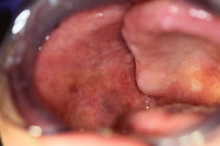
Palatal leukoplakia
Extra-oral exam was unremarkable. Intra-oral exam revealed leukoplakias on the lower left alveolar ridge and the palatal mucosa. Subtle lesions were noticed on the right and left buccal mucosa as well. This presentation is most consistent with proliferative verrucous leukoplakia. I recommended biopsies of the most prominent lesions. Their diagnosis ranged between epithelial atypia and mild epithelial dyplasia. The patient was kept on strict 3-6 month follow-ups. New lesions and previous lesions undergoing changes in shape, size and texture were biopsied. We still see her periodically in clinic. 

Proliferative Verrucous Leukoplakia:

Proliferative verrucous leukoplakia (PVL) is a condition characterized by development of multiple leukoplakias in the oral cavity. The lesions may be subtle at first but they gradually evolve into more prominent plaques over a period of time. These leukoplakias have the ability to transform into verrucous carcinoma or squamous cell carcinoma. 

Unfortunately, we cannot treat this condition. Complete excision is also not a viable option because usually the entire mucosa is involved and because of the high rate of recurrence. The only thing we can do is monitor for changes. For that, it is recommended that patients with PVL are seen are regular 3-6 month intervals. Any changes in the lesions is documented and lesions showing changes in shape, size and texture are biopsied immediately. If the biopsy result grades a lesion between epithelial atypia and mild epithelial dysplasia, then the lesion will continue to be monitored. If the lesion is graded between moderate epithelial dysplasia and carcinoma in situ, it is mandatory to perform complete excision of that lesion. 

The patient will require monitoring for the rest of his life. For patient's that don't return for their periodic follow-ups, rates of transformation is high. 


Tuesday, January 27, 2015

The Case of a White Lesion

A 48-year-old female presented with a white lesion on her lower labial mucosa noted during her follow-up exam in the implant clinic. The lesion was asymptomatic and the patient was unaware of it presence. The hygienist evaluating this patient recorded the dimensions as 40 X 40 mm. She also documented in her noted that the lesion appeared to be "well defined". (Hygienists take wonderfully thorough notes!). There was no significant medical or social history.
Leukoplakia in a 48-year-old female

The lesion was diagnosed as leukoplakia and an excisional biopsy was recommended. The biopsy result read mild epithelial dysplasia. The patient was kept under close observation because almost a third of these tend to return. 

Leukoplakia:

The traditional definition of leukoplakia lumps up all unclassifiable white lesions with no identifiable etiology. I was taught that leukoplakias are white lesions with a crisp margin, i.e margins that you can trace out using a pen. The rationale behind this odd thought is that it is humanly impossible to irritate the same number of mucosal cells each time. You will irritate some cells at first, a little more the next time, and a little less next time after that. This will result in a gradient, from the normal skin to a little white, more white, less white and normal skin and "not a crisply defined margin". A crisply defined margin forms when the cells constituting that margin are dysplastic.

Leukoplakias need to undergo biopsies to establish their histologic grade. Smaller ones can undergo excisional biopsies while the larger ones can be incisionally biopsied. Biopsies resulting in a diagnosis of epithelial atypia or mild epithelial dysplasia should be kept under close observation. Recurrent lesions or changes in shape, size or texture of residual lesions should be promptly biopsied and treated accordingly.

For lesions graded as moderate to severe epithelial dysplasia, or carcinoma in situ, surgical excision of the involved tissue is recommended. These patients are also kept under close observation to monitor for recurrent lesions.

There have been numerous attempts to find non-invasive cures/treatments of leukoplakia, as of now, very little evidence supports their use. 

Monday, January 26, 2015

The Case of "Brown-Brown Thing"

My 3-year-old nephew pointed out something on my 64-year-old mother's hand the other day, and asked her what that brown-brown thing was. I asked my mother if he had located an age-spot (lentigines). She told that she didn't think so because this one felt slightly elevated, kind of "stuck on". Unfortunately, her lesion was about 2 X 2 mm in size and kind of hard to photograph, but I do have another really nice picture of this process.

Seborrheic keratosis

The lesion on my mother's hand was seborrheic keratosis, a benign proliferation of skin cells that is common in individuals over the age of 40. It presents as a brown, slightly raised and well defined plaque with an irregular surface. The appearance is described as either "stuck on" or as "wax drippings". Face, trunk, arms and legs are commonly affected. Usually more than one lesion is present. 

Removal by cryotherapy or surgery can be performed for esthetic purposes. 

In rare instances, an internal malignancy can lead to formation of numerous lesions of itchy seborrheic keratosis. This is known as the Leser-Trelat sign.  

Sunday, January 25, 2015

The Case of White Striations

I was asked a review some glass slides for a case last week. The patient was a 48-year-old woman who had reported to the oral surgery clinic with a complaint of painful bilateral lesions on the buccal mucosa. The lesions had been present for over 3 years. They were especially painful when the patient tried to consume something acidic or spicy. The history was waxing and waning of the lesions was not provided. 

A metal crown, placed several years ago was present adjacent to one of the lesions. The patient was hypertensive and diabetic. She was currently taking medications for both these diseases.


48-year-old woman with erosive lichen planus. The white striae can be identified at the margins of the ulcers.
Courtesy: Dr. Sidra-tul-Muntaha
The lesions were biopsied by the surgeon and submitted for histopathologic examination. The general pathologist signed this process out as chronic mucositis. The surgeon then asked me to review the slides. She told me that the striations were so prominent that it could only be lichen planus or a lichenoid reaction.

And she was right. The history, the clinical picture and the histopathologic presentation all favored a lichenoid process. Since this presentation was bilateral, lichen planus was the most appropriate diagnosis.

Lichen Planus:

Lichen planus is an autoimmune process characterized by presence of white striae. It has two major forms, reticular and erosive. Reticular lichen planus is asymptomatic and usually discovered during routine oral exams. It appears as symmetrical and bilateral, white lace-like pattern on the patient's buccal mucosa. Occasionally other sites may also be involved. It does have the ability to evolve into the more painful, erosive form. Erosive lichen planus in painful because of mucosal atrophy and ulcer formation. This type of lichen planus is associated with desquamative gingivitis.

Like all autoimmune processes, lichen planus waxes and wanes over a period of time. A flare is noted every few weeks to months.

No management is required for the reticular type.

For erosive lichen planus, potent topical steroids should be applied 4-6 times a day to control the flare. Once the lesions heal up, the patient can stop drug application until the next flare.

It is important to keep in mind that steroid application inside the mouth may lead to candidiasis. In such instances, the steroid will no longer heal the lesions and the symptoms would continue. Following confirmation of a candida infection by culture or cytology, a course of appropriate anti fungal therapy is recommended. I prefer using Clotrimazole troches for this purpose. Fifty tablets are dissolved slowly in mouth over a course of 10 days. Once the candida infection in under control, steroid application can begin again.

Note:

This patient is particularly interesting because she had a metal crown adjacent to one of the lesion, and she was using both antihypertensive and hypoglycemic drugs. All three factors have been linked to lichenoid reactions. In this case, these processes were easy to rule out because this woman's presentation was symmetric and bilateral. Lichenoid reactions are "always" asymmetric or unilateral.

A special thanks to Dr. Sidra-tul-Muntaha for sharing this case.

Friday, January 23, 2015

The Case of a Black Spot on the Lips

A student in the screening clinic had documented the lesion seen in the picture as a mole. 


Melanotic macule in a 40-year-old male
The brown spot seen on this 40-year-old man's lip is called a melanotic macule

Melanotic Macule:

Melanotic macule is a focal discoloration of the oral mucosa. It is believed to be a result of an increased expression of melanin (the pigment that gives color to our skins). The color varies from brown to black. The cause for development of melanotic macules is not known. 

Treatment is usually not required. If a patient wishes to remove it for cosmetic reasons, conservative excision is sufficient. 

It is important that pigmented lesions on the maxillary gingiva and palate are kept under close observation or excised and submitted for histopathologic examination. This is because of the high incidence of oral melanoma at these sites. 

Thursday, January 22, 2015

The Case of Lump on Tongue

A very sweet 70 something year-old woman came through the clinic. She was extremely concerned that she has oral cancer. She stuck out her tongue and showed me the small bump on mid dorsal.

70-year-old female with a fibroma

She was unable to confirm a history of trauma in the area but did add that her memory had not been reliable recently. 

The bump was firm on palpation. Following the oral exam, I put two things in my differential. The first one was a fibroma, because that's what this bump looked and felt like. The other one was granular cell tumor, because dorsal tongue is a frequent site of occurrence and I really wanted a good clinical picture for that tumor.

I assured the patient that I would be extremely shocked if this ended up being cancer and recommended an excisional biopsy. The biopsy results confirmed the diagnosis of a fibroma

Fibroma:

Fibromas are extremely common, benign tumors commonly found in the oral cavity. They are believed to be an exaggerated response to trauma. The traumatic event leads to excess deposition of collagen fibers in the area resulting in a small bump or lump. 

Conservative excision is the preferred form of treatment.

Wednesday, January 21, 2015

The Case of "Suspected" Plasma Cell Gingivitis

A resident in the general practice told me that she had a patient in her chair whose gingiva looked identical to the picture of plasma cell gingivitis in Dr. Allen's book*. The patient was 23-year-old intellectually disable woman. Her parents told us that her gums are usually this red. They schedule cleanings under sedation every six months because she refuses to brush. They couldn't recall if it gets better after the cleaning appointment.


23-year-old diabetic female with plaque deposition and gingivitis
(I apologize for the quality of the picture, the girl was wonderful during the exam but would not let us use retractors)

The patient had a complex medical history with all sorts of problems including heart disease and diabetes.

Based on the medical history and the clinical picture, this condition was diagnosed as diabetic gingivitis. (Plasma cell gingivitis is an entirely different process, and hopefully will be discussed in the future).


Diabetic gingivitis is not a unique entity, it is just a more severe presentation of "regular" gingivitis seen in diabetic people. It is believed that insufficient glucose control leads to increased bacterial growth and decreased resistance to infection. Patients with diabetes need to be more vigilant about their hygiene routines. Brushing twice a day, flossing daily and professional cleanings every six months are mandatory for this purpose. 


Unfortunately, this young woman was not capable of maintaining such a thorough cleaning regimen but she had minimal dental caries and her periodontal health was not significantly bad. The every 6 month cleaning regimen appeared to be working well for her. This is why it was extremely difficult for me to understand why her regular dentist had recommended complete extractions for this 23-year-old. I asked the parents if she was in pain, they said no. I asked them if she had trouble eating, they said no. "Then why?" was my final question. Their answer was because she didn't brush. 


I spent the next half an hour with her mother and father explaining to them why, in my opinion, full extractions on someone who already has a very difficult life is not a good idea. I don't know if I had managed to convince them and honestly, I am too afraid to ask.

* Oral and Maxillofacial Pathology - Neville, Damm, Allen and Bouqout